
![]() Eliminates Subjectivity: Provides unbiased, numerical data on visual acuity and contrast sensitivity, bypassing limitations of manual observation.
Eliminates Subjectivity: Provides unbiased, numerical data on visual acuity and contrast sensitivity, bypassing limitations of manual observation.
![]() Reproducible Results: Automated measurement and analysis ensures high consistency across experiments.
Reproducible Results: Automated measurement and analysis ensures high consistency across experiments.
![]() Hands-Free Operation: System automatically adjusts stimulus based on real-time head tracking, requiring no manual intervention.
Hands-Free Operation: System automatically adjusts stimulus based on real-time head tracking, requiring no manual intervention.
![]() No Specialized Training Needed: Designed for ease of use, making it practical for a wide range of researchers.
No Specialized Training Needed: Designed for ease of use, making it practical for a wide range of researchers.
![]() Stress-Minimized Testing: Assesses vision without physical restraint, sedation, need for surgery promoting natural behavior.
Stress-Minimized Testing: Assesses vision without physical restraint, sedation, need for surgery promoting natural behavior.
![]() Leverages Natural Reflex: Based on the innate optomotor response, eliminating need for animal training.
Leverages Natural Reflex: Based on the innate optomotor response, eliminating need for animal training.
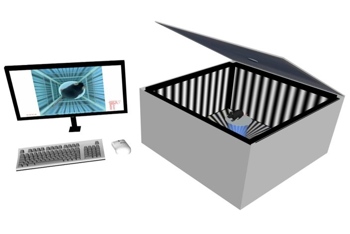
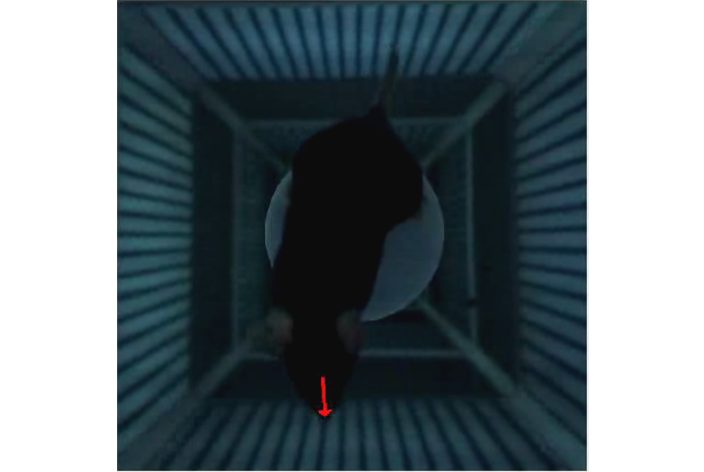
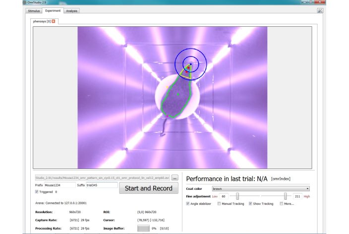
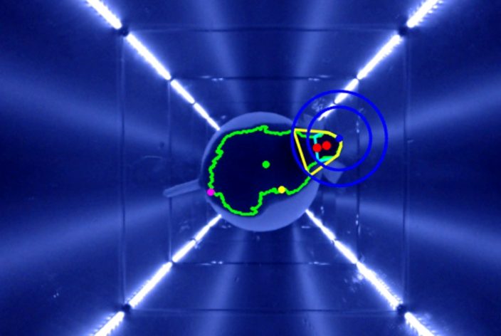
![]() Continuous Alignment: Virtual stimulation cylinder continuously aligns with the animal’s head for accurate data collection.
Continuous Alignment: Virtual stimulation cylinder continuously aligns with the animal’s head for accurate data collection.
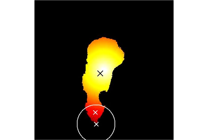
![]() Synchronized Head Movement: Precisely evaluates head movements synchronous to stimulation for quantitative OMR.
Synchronized Head Movement: Precisely evaluates head movements synchronous to stimulation for quantitative OMR.

Characterization of Vision
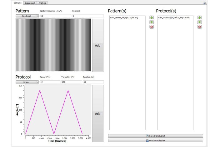
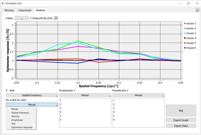
Objectively measure visual acuity, contrast sensitivity alongwith spectral and temporal sensitivity.

Screening for Vision Defects
Identify and quantify vision impairments.

Tracking Disease Progression & Recovery
Characterization or pre-clinical testing in relevant disease models like glaucoma, or other ocular diseases.

Phenotyping New Breed Lines
Rapidly assess the visual capabilities of novel mouse strains.

Quantification of Treatment Response
Evaluate the efficacy of therapeutic interventions on visual function.