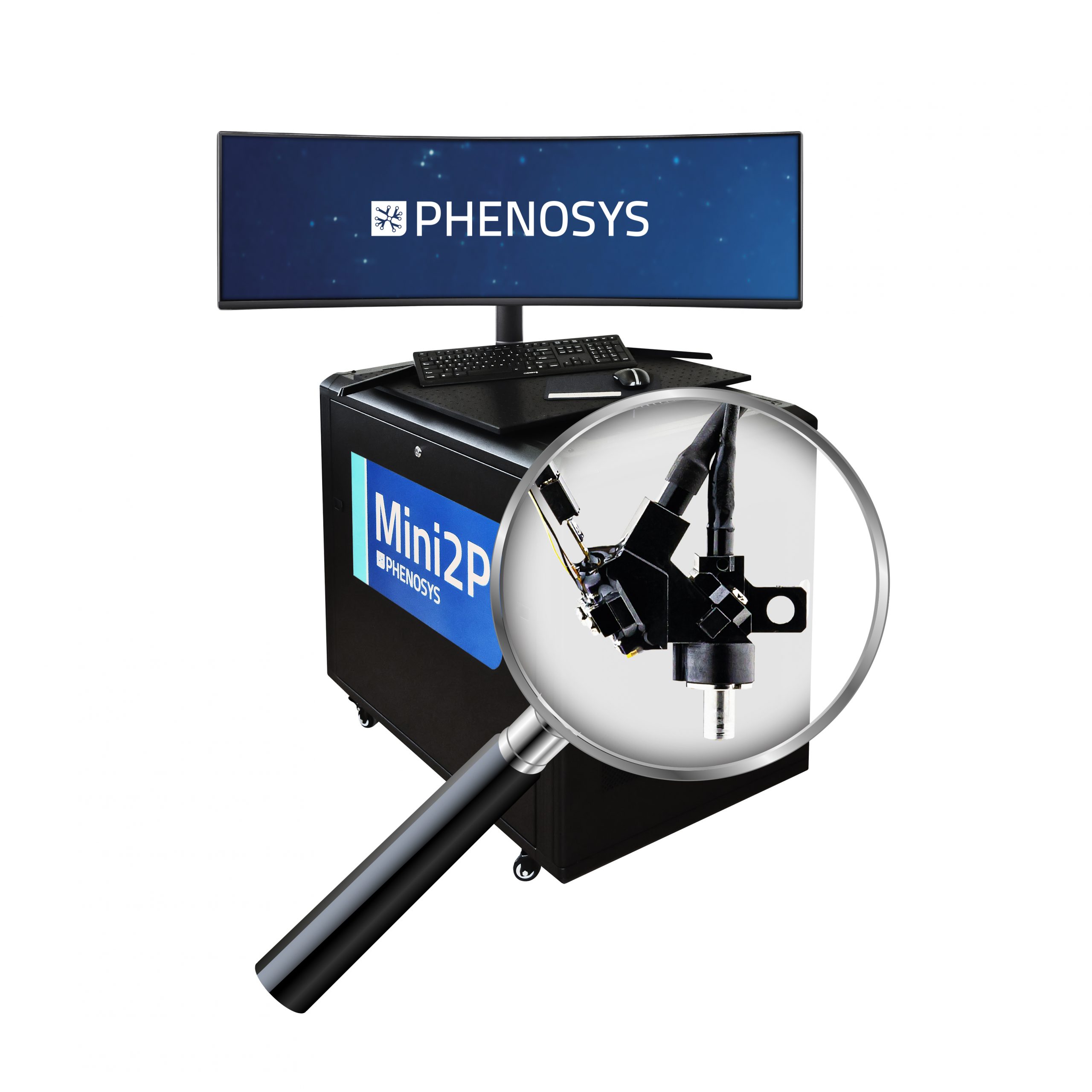
Mini2P: The Complete System

The Mini2P is a miniature two-photon microscope for fast, high-resolution, multi-plane calcium imaging in freely moving mice. Weighing under 3 g with a highly flexible connection cable, Mini2P enables stable imaging without hindering animal behaviour. Its optimized optical system allows stable simultaneous recordings of neuronal activity of more than thousand cells in different brain regions.

PhenoSys presents this innovative technology as a turnkey solution, including the miniature 2P microscope with its fiber optics, laser, detection unit and flexible DAQ hardware and software. Get your complete system and have it ready to run within hours.

The Mini2P technology originates from the Kavli Institute in Norway (Zong W., Obenhaus HA., Skytoen ER., Eneqvist H., de Jung NL., Vale R., Jorge MR., Moser M., and Moser El., 2022, Cell 185, 1240-1256).

  • Head-staged miniature two-photon microscope for calcium imaging in freely moving animals, developed by Zong et al. (1).
  • Total solution suite:
    • Ultra-light microscope body (2.4g) with MEMS scanner for X-Y-scanning and µTlens for Z-scanning.
    • Two photon laser (920nm) with clean femtosecond pulses (<100 fs).
    • Light and flexible fiber optics and cable, all at a length of 2.2 m for maximal freedom to move for the animal.
    • Detection unit with two high-sensitive GaAsP-type photomultipliers.
    • DAQ-BNC interface board for flexible integration/synchronization of/with additional devices.
    • DAQ computer with preinstalled and configured Matlab-based software (ScanImage).
    • Compact hardware design, everything integrated on a mobile trolley.
    • The package comes ready-to-use.
    • Full technical support
  • Fast imaging (15 Hz for 256×256 pixels) in a large field of view 500×500 µm2.
  • Multilayer imaging into 240 µm depth.
  • Maximum resolution of 1.3 µm x 1.3 µm x 15 µm.
  • Set of interchangeable objectives to image e.g. visual cortex, medial entorhinal cortex or hippocampus.
  • Simultaneous activity imaging of more than 1,000 neurons in one region or recoding of more than 10,000 cells in adjacent field-of-views.
  • Stable high-quality imaging across a wide spectrum of mouse behaviours, including instances such as climbing and jumping.
  • Raw data is fully accessible and the system is individually extendable.


(1) Zong W., Obenhaus HA., Skytoen ER., Eneqvist H., de Jung NL., Vale R., Jorge MR., Moser M., and Moser El., 2022, Cell 185, 1240-1256

  • Single wavelength fiber laser: 920 nm, <100 fsec
  • Mini2P microscopy body:
    • Weight: 2.4 g
    • Quartet of microtunable lenses (µTlens) for z-scanning with 240 µm depth
    • MEMS mirror with 15 Hz frame rate at 256 x 256 pixels for a field of view of 500 µm x 500 µm
    • Resolution: 1.3 µm x 1.3 µm x 15 µm
    • Base plate and stitching adapter (for FOVs of up to 4 mm2)
  • Interchangeable objectives:
    • D0213 (with cover glass to image e.g. visual cortex)
    • D0254 (with GRIN lens to image e.g. hippocampal CA1)
    • D0277 (with prism to image e.g. medial entorhinal cortex)
  • Excitation fiber:
    • Length: 2.2 m
  • Collection fiber:
    • Length: 2.5 m
  • Cable Harness:
    • 6-wire bundle to control µTlens and MEMS mirror
    • Length: 2.5 m

The Mini2P is a miniature 2-photon microscope brain explorer for fast high-resolution calcium imaging in freely-moving mice.

The Mini2P has a weight of only 2.4 g.

The Mini2P technology originates from the Kavli Institute in Norway (1).

(1) Zong W., Obenhaus HA., Skytoen ER., Eneqvist H., de Jung NL., Vale R., Jorge MR., Moser M., and Moser El., 2022, Cell 185, 1240-1256

We supply the Mini2P together with a mobile cart containing the DAQ interface and the laser.

The Mini2P originates from the Kavli Institute, Norway: Mini2p website


Zong W., Obenhaus HA., Skytoen ER., Eneqvist H., de Jung NL., Vale R., Jorge MR., Moser M., and Moser El., 2022, Cell 185, 1240-1256