The optomotor response (OMR) is a reflex used to assess visual function. The PhenoSys qOMR (quantitative optomotor response) is a unique system that objectively measures the OMR with minimal experimenter effort. It employs a virtual stimulation cylinder that continuously aligns with the animal´s head position. Based on real-time head tracking, quantitative OMR measurements automatically provide visual acuity and contrast sensitivity. This is a PhenoSys Collaboration product brought to market together with its developer, Dr. Friedrich Kretschmer. New:qOMR-XT for rats
Check out this video to see how it is operated.
Hardware features
Software features
General application areas:
Characterisation or preclinical testing in relevant disease models, for example:
Investigation of various aspects of vision in mice and other rodents:
The PhenoSys qOMR is a PhenoSys Collaboration product. These products are brought to market together with the scientists who developed them.
qOMR is a joint product of Dr. Friedrich Kretschmer and PhenoSys.
qOMR is based on his publications:
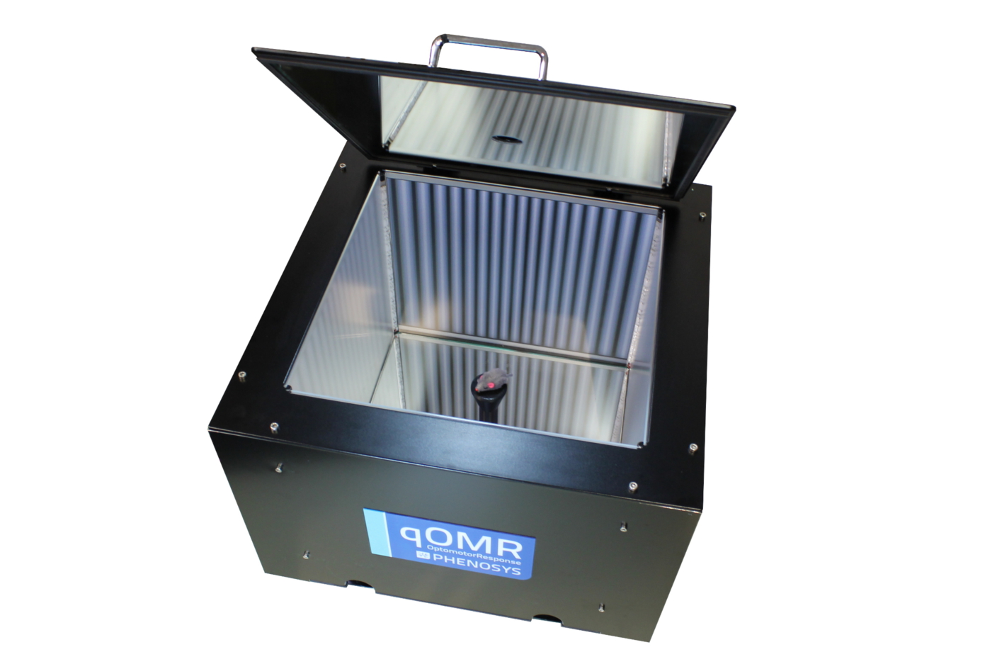
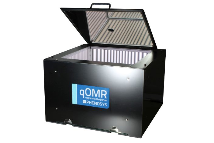
PhenoSys qOMR main unit with partially opened lid.
Hardware Features
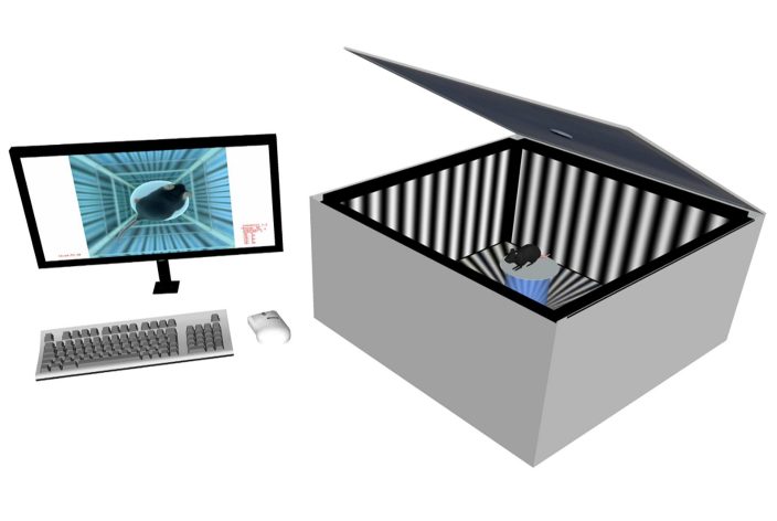
Principle of the experiment with a mouse on top of the platform. The stimulus is normally invisible to the IR camera.
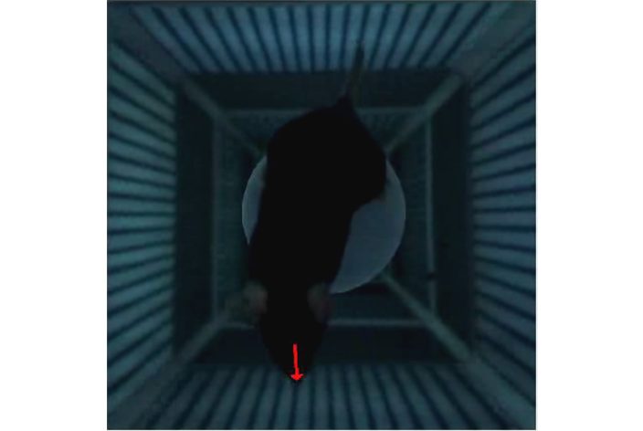
Video-based real-time tracking of head movement. The contour and pointing direction is determined by a fast algorithm using minimal assumptions for the geometry of the animal.
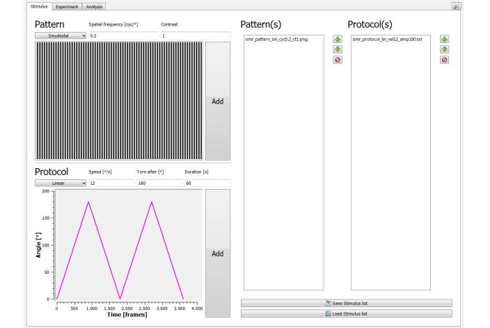
Software omrStudio: stimulus design specifying the presented pattern and the movement scheme
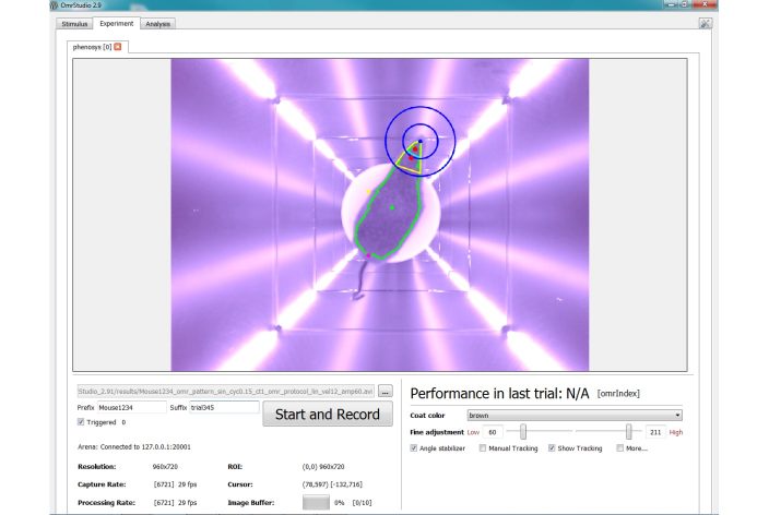
Software omrStudio: running experiment.
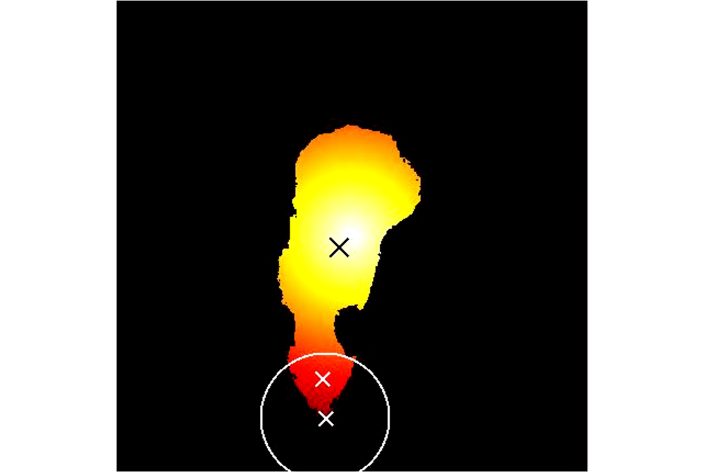
Actual measurement data (single frame) and recorded video with superimposed tracking results:
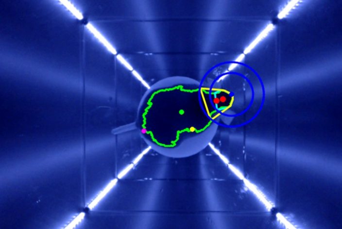
qOMR video with visible stimulus.
The visibility of the stimulus is enhanced compared to the normal qOMR for better clarity.
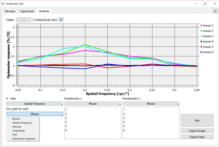
Software omrStudio: data analysis.
Kretschmer V, Schneider S, Matthiessen PA, Reichert D, Hotaling N, Glasßer G, Lieberwirth I, Bharti K, De Cegli R, Conte I, Nandrot EF, May-Simera HL. Deletion of IFT20 exclusively in the RPE ablates primary cilia and leads to retinal degeneration. PLoS Biol. 2023 Dec 4;21(12):e3002402. doi: 10.1371/journal.pbio.3002402. Epub ahead of print. PMID: 38048369.
Liu Y, Li Q, Yan T, Chen H, Wang J, Wang Y, Yang Y, Xiang L, Chi Z, Ren K, Lin B, Lin G, Li J, Liu Y, Gu F. Adenine base editor-mediated splicing remodeling activates non-canonical splice sites. J Biol Chem. 2023 Nov 8:105442. doi: 10.1016/j.jbc.2023.105442. Epub ahead of print. PMID: 37949222.
Huh, C.Y.L., Leinonen, H., Nakayama, T., Tomasello, J.R., … & Gandhi, S.P. (2022). Retinoid therapy restores eye-specific cortical responses in adult mice with retinal degeneration. Current Biology.
Suh, S., Choi, E. H., Leinonen, H., Foik, A. T., Newby, G. A., Yeh, W. H., … & Palczewski, K. (2020). Restoration of visual function in adult mice with an inherited retinal disease via adenine base editing. Nature biomedical engineering, 1-10.
Lees, R. N., Akbar, A. F., & Badea, T. C. (2020). Retinal Ganglion Cell defects cause decision shifts in visually evoked defense responses. Journal of Neurophysiology 124:5,1530-1549
Chan, K., Hoon, M., Pattnaik, B. R., Ver Hoeve, J. N., Wahlgren, B., Gloe, S., … & Jansen, E. (2020). Induced Retinal Functional Alterations and Second-Order Neuron Plasticity in C57BL/6J Mice. Investigative Ophthalmology & Visual Science, 61(2), 17-17.
Thomson, B. R., Grannonico, M., Liu, F., Liu, M., Mendapara, P., Xu, Y., … & Quaggin, S. E. (2020). Angiopoietin-1 Knockout Mice as a Genetic Model of Open-Angle Glaucoma. Translational Vision Science & Technology, 9(4), 16-16.
Kretschmer, V., Patnaik, S. R., Kretschmer, F., Chawda, M. M., Hernandez-Hernandez, V., & May-Simera, H. L. (2019). Progressive characterization of visual phenotype in Bardet-Biedl syndrome mutant mice. Investigative ophthalmology & visual science, 60(4), 1132-1143.
Wang, X., Zhao, L., Zhang, J., Fariss, R. N., Ma, W., Kretschmer, F., … & Gan, W. B. (2016). Requirement for microglia for the maintenance of synaptic function and integrity in the mature retina. Journal of Neuroscience, 36(9), 2827-2842.
Kretschmer, F., Tariq, M., Chatila, W., Wu, B., & Badea, T. C. (2017). Comparison of optomotor and optokinetic reflexes in mice. Journal of neurophysiology, 118(1), 300-316.